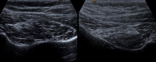
Fig. 2.
Ultrasonographic appearance of the infraspinatus muscle. The muscle without fatty degeneration (left) shows a clear central tendon and hypoechoic surrounding muscle. The muscle with fatty degeneration (right) shows increased echogenicity at the surrounding muscle with a blurred central tendon.

