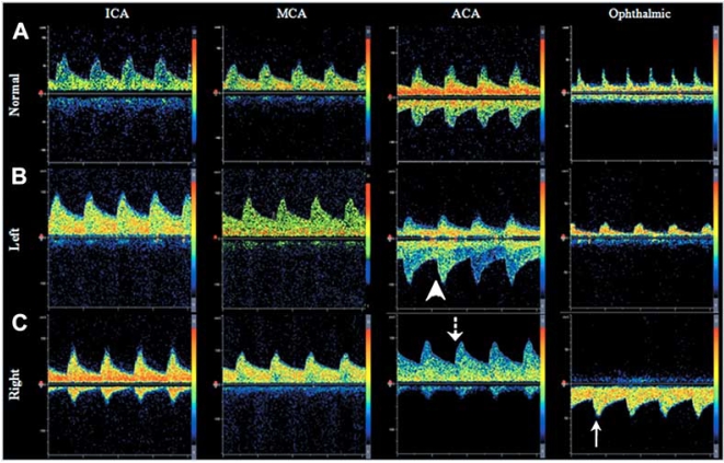
Fig. (4). Transcranial Doppler Ultrasound demonstrating patterns of collateral circulation.
Panel A. Normal flow velocities and direction of flow in the internal (ICA), middle (MCA), and anterior cerebral arteries (ACA), and ophthalmic artery. Panels B and C. Example of recruitment of collateral circulation through the anterior cerebral artery (Panel B) and ophthalmic artery (Panel C) in a patient with right internal carotid occlusion below the level of the ophthalmic artery origin. Note that there is inversion of flow direction in the ACA (dotted arrow), compared to the normal side (arrow head). Panel C shows retrograde flow through the ophthalmic artery (solid arrow), demonstrating collateral circulation.

