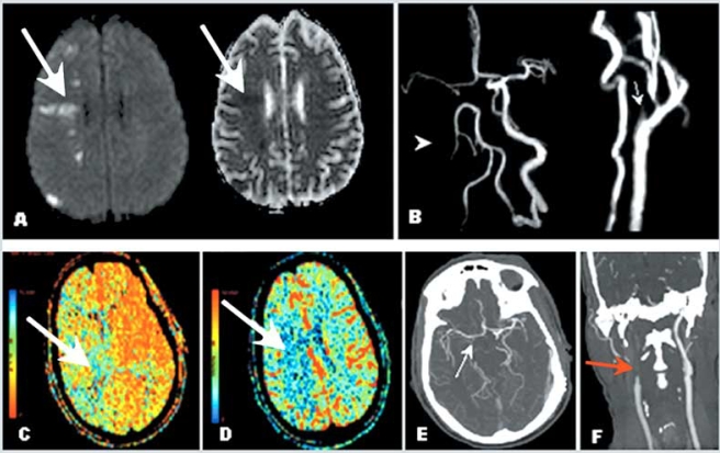
Fig. (7). Example of patient with carotid artery occlusion and clinical fluctuations related to hemodynamic factors; evaluation of collateral circulation and cerebral perfusion.
A 56-year-old right-handed man presented with confusion and left side weakness. He was found to have complete occlusion of the right internal carotid artery. Brain MRI demonstrated small infarctions in borderzone territories, located between anterior and middle cerebral artery, and between posterior and middle cerebral artery territories. Panel A demonstrates bright signal in the right hemisphere (Diffusion Weighted Imaging) and corresponding dark signal (Apparent Diffusion Coefficient, right of panel A) consistent with acute infarction. Magnetic Resonance Angiography (Panel B) and Computed Tomography Angiography of the neck and head (Panel F) confirmed occlusion of the proximal internal carotid artery after its origin (arrows). Panels B and E demonstrate lack of visualization of the right internal carotid artery intracranially as well as collateral flow through the anterior communicating artery suggested by visualization of the middle cerebral artery ipsilateral to the carotid occlusion. The patient presented severe worsening of left side weakness when anti hypertension medications were started, and developed left hemiplegia. A perfusion study (CTP) showed prolonged meant transit time (Panel C) and normal cerebral blood volume in the right middle cerebral artery territory (Panel D), consistent with a mismatch (ischemic penumbra). Repeat brain MRI showed no extension of the infarction (not shown). Collateral flow failure was suspected and blood pressure was elevated with intravenous fluids. The patient presented marked improvement and remained stable thereafter. He was discharged to rehabilitation, and at follow up 2 months later, had minimal left hemiparesis, was able to ambulate without supportive device and able to use fully his left hand. Modified Rankin Scale score was 1.

