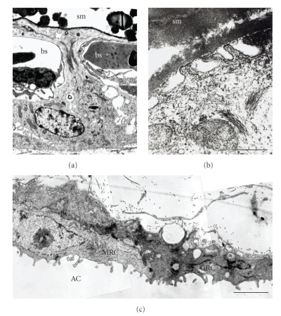
Figure 4.
(a) A CC cell shows its typical cytoplasmic lateral projections which separate the shell membrane (sm) from the capillaries of the blood sinus (bs). Original magnification: 11,200x; scale bar: 2 μm. (b) Apical pits are formed by CC cell membrane adjacent to the inner shell membrane (sm). Original magnification: 70,000x; scale bar: 0.5 μm. (c) Basal cells (BC), mitochondria-rich cells (MRC), and granule-rich cells (GRC) characterize the CAM allantoic epithelium which faces the allantoic cavity (AC). Original magnification: 21,700x; scale bar: 1 μm. M. G. Gabrielli, unpublished.

