Figure 5.
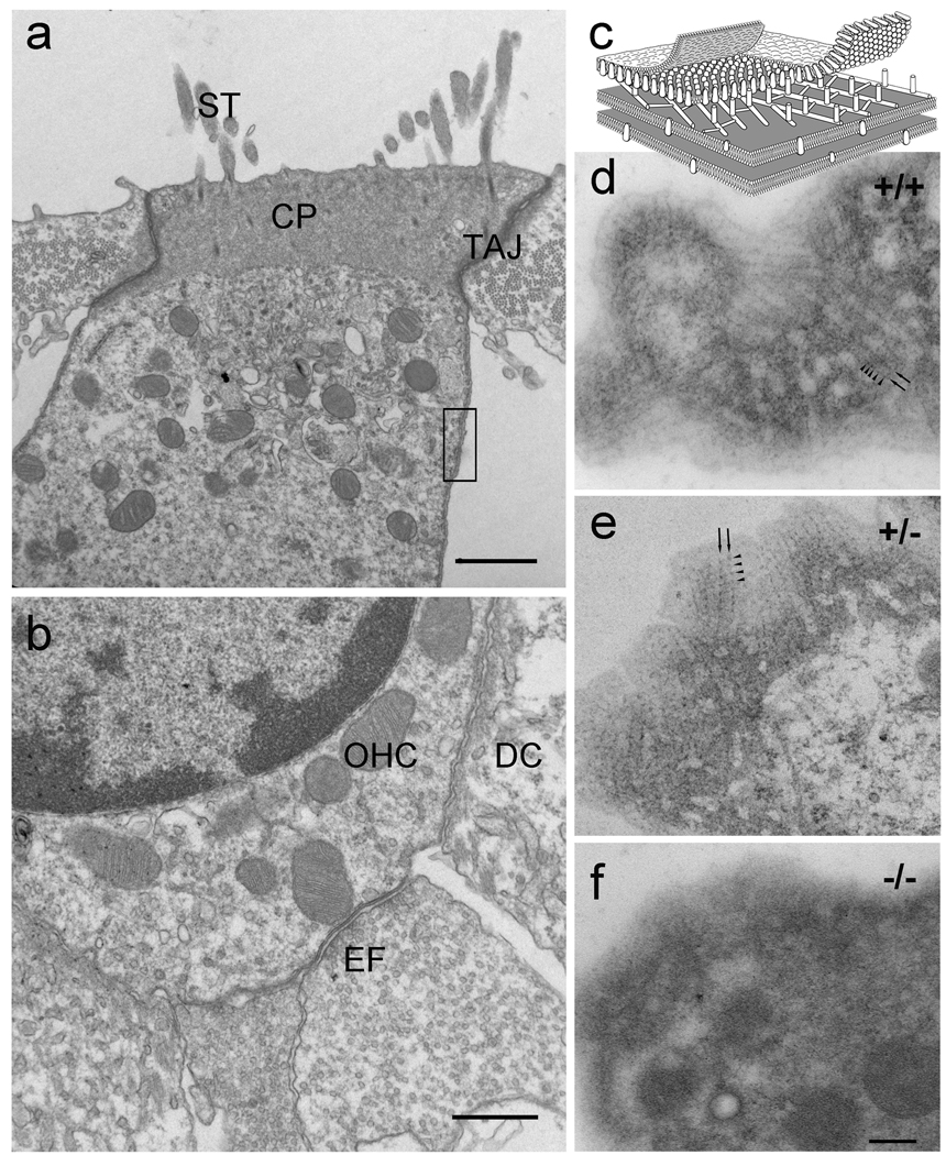
Disruption of the cortical lattice in prestin-null OHCs. At the electron microscopic level the prestin-null OHC shows the characteristic organization of a typical OHC (a, b) with stereocilia (ST), cuticular plate (CP), tight-adherens junction (TAJ), subsurface cisternae underlying the lateral plasma membrane, attachment to the Deiters’ cell (DC), and efferent synapses (EF). (c) Schematic diagram of a portion of the lateral wall of the OHC (equivalent to the region delineated by the rectangle in a) showing the cortical actin-spectrin cytoskeletal lattice between the lateral plasma membrane and the underlying subsurface cisterna. The parallel actin filaments (arrows) and the periodic puncta (arrowheads) characteristic of the cortical actin-spectrin lattice are clearly visualized in thin section grazing or tangent to the curved surface of the OHC lateral wall (d, e) from the WT and hetereozygote but are absent in the prestin-null OHC (f). Magnification bars equal: 1 µm in a; 0.5 µm in b; and 200 nm in d–f.

