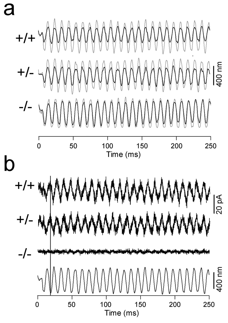
Figure 6.
Changes in axial stiffness and piezoelectric properties in prestin-deficient OHCs. (a) Measurement of passive axial stiffness. Examples of free-fiber (in light-color lines) and loaded-fiber motions (heavy lines) measured from the OHCs isolated from the cochleae of WT, heterozygous, and homozygous mice. The axial stiffness for the cells is 3.1 (+/+), 2.6 (+/−), and 0.9 (−/−) mN/m. (b) Examples of compression-evoked charge movement. Compression of the cell by a loaded fiber evoked an outward current in both +/+ and +/− OHCs. The current was absent in prestin-null OHCs. Bottom panel shows the waveform of the fiber motion. The black line indicates the direction of fiber motion (compression) and the outward current.

