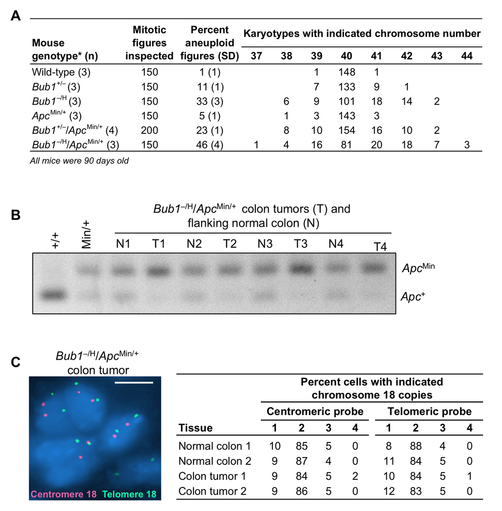
Figure 5. Apc LOH in colon tumors from Bub1−/H/ApcMin/+ mice.
(A) Chromosome counts on splenocytes of 3-month-old wild-type, Bub1+/−, Bub1−/H, ApcMin/+, Bub1+/−/ApcMin/+ and Bub1−/H/ApcMin/+ mice.
(B) PCR-based screening for Apc LOH in colon tumors from Bub1−/H/ApcMin/+. Tail DNA samples from wild-type and ApcMin/+ mice were used as PCR controls. Note that colon tumors from Bub1−/H/ApcMin/+ mice consistently show preferential amplification of the ApcMin allele, indicating loss of the Apc+ allele.
(C) Interphase FISH image of a 5 µm colon tumor section of a Bub1−/H/ApcMin/+ mouse hybridized to both centromeric (red) and telomeric (green) chromosome probes. Quantification of the chromosome 18 copies in colon cancer cells and flanking normal colon epithelial cells. FISH signals of one hundred cells were counted per section. Scale bar is 5 µm.

