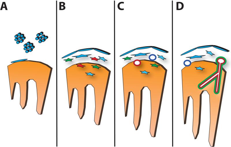
Figure 1. Steps in Coronary Vascular Development.
In (A), the epicardium is formed by migration of proepicardial cells (blue) to the surface of the heart tube (orange), forming an epithelial sheet. In (B), epithelial-to-mesenchymal transformation produces cells with hemangioblast (red), smooth muscle (green) and fibroblast (blue) cell fates. In (C), a primitive endothelial plexus forms from the endothelial progenitors, with venous-fated endothelial tubes (dark blue) forming subepicardially and arterial-fated endothelial tubules forming intramyocardially (red). In (D), the endothelial plexus remodels and recruits vascular smooth muscle and other adventitial cells to form the mature coronary arteries (red) and veins (dark blue).

