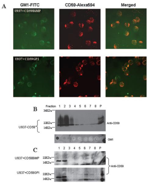
FIGURE 5.
Incorporation of BiMP CD59 and GPI-anchored CD59 into U937 cells. A, Cocapping is illustrated by immunofluorescence analysis of CD59 colocalization with the cholera toxin-ganglioside GM1 raft protein. Magnification is ×400. Sucrose gradient ultracentrifugation of the stable CD59-expressing cell line (B) and incorporated CD59 lines (conducted as described in Materials and Methods) (C) confirm the localization of CD59 in the detergent insoluble (lipid raft) fractions. The nuclear pellet (P) was resuspended by brief sonication on ice. Fractions containing GM1 were identified by dot blotting to confirm the location of lipid rafts in the gradient (B).

