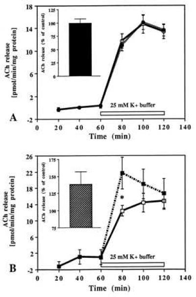
Figure 7.

Time-course effects of IGF-I (A) and IGF-II (B) on evoked ACh release from slices of the striatum. Tissue slices were stimulated with 25 mM K+ buffer in the presence (dotted lines) or absence (solid lines) of 10−8 M IGF-I and 10−10 M IGF-II. Evoked ACh release was unaffected by IGF-I (A) while potentiated significantly at the early phase of stimulation by IGF-II (B). A and B (Insets) represent total release as percentage of control for the respective peptides over 60 min stimulation period. Data are expressed as the mean ± SEM (n = 10–12). *P < 0.05.
