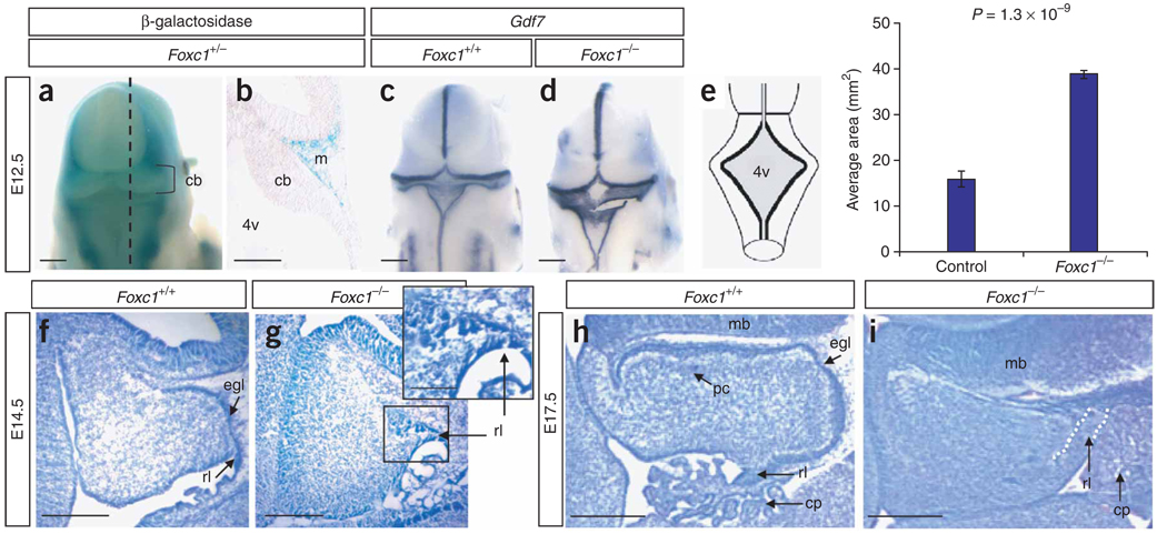
Figure 3.
Morphological cerebellar phenotype of Foxc1 −/− embryos. (a) Dorsal view of the head from an X-gal–stained Foxc1 +/− (Mf1 +/LacZ) embryo at E12.5 showing that Foxc1 expression excludes the developing brain. (b) Midsagittal section through embryo in a (dashed line) showing expression in the mesenchyme (m) adjacent to the cerebellar anlage (cb). (c) Dorsal view of Gdf7 expression in a wild-type embryo. (d) Dorsal view of Gdf7 expression in a Foxc1 −/− embryo revealing an enlarged fourth ventricle roof plate (4v). (e) Schematic of the dorsal view of an E12.5 embryo depicting the 4v area (shaded gray) measured in Foxc1 −/− and littermates. The 4v was 2.5-fold larger in Foxc1 −/− embryos. (f) Cresyl violet–stained midsagittal section of the developing cerebellar anlage in a wild-type E14.5 embryo. (g) Midsagittal section of the developing cerebellar anlage in a Foxc1 −/− E14.5 embryo showing that the rhombic lip (rl) and external granule cell layer (egl) are abnormal and highly disorganized. (h) Midsagittal section through the cerebellar vermis of a wild-type E17.5 embryo showing distinct egl and Purkinje cell (pc) layers. (i) Midsagittal section through the cerebellar vermis of a Foxc1 −/− E17.5 embryo showing a lack of distinct egl and pc layers, an abnormal rl and an expansion of the choroid plexus (cp). Scale bars, 100 (a,c,d), 50 (b,f–i) or 20 µm (g inset).

