Figure 5.
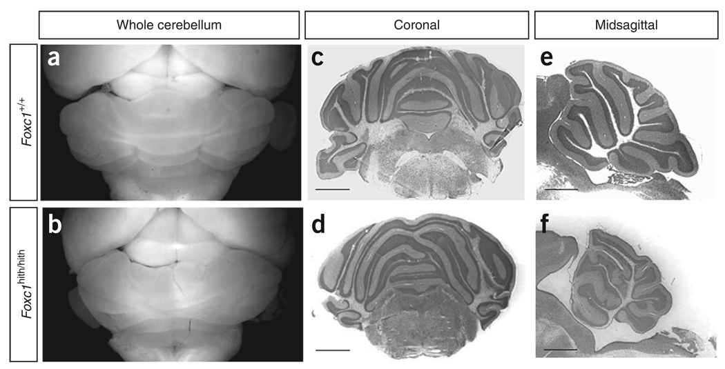
Cerebellar morphology of Foxc1 hith/hith mice. (a,b) Dorsal views of mouse brains at postnatal day (P) 30. Mean cerebellar area between Foxc1 hith/hith (5.63 arbitrary units (a.u.) ± 0.31 s.e.m.) and littermates (8.60 a.u. ± 0.03 s.e.m.) reveals significant hypoplasia in these adult mutants (P< 0.01). (c,d) Cresyl violet–stained coronal sections through the cerebellum. (e,f) Cresyl violet–stained midsagittal sections through the vermis of brains of mice at P30. The wild-type cerebellum (a) has a stereotypic foliation pattern, which is disrupted in the homozygous mutants. Scale bars, 200 µm.

