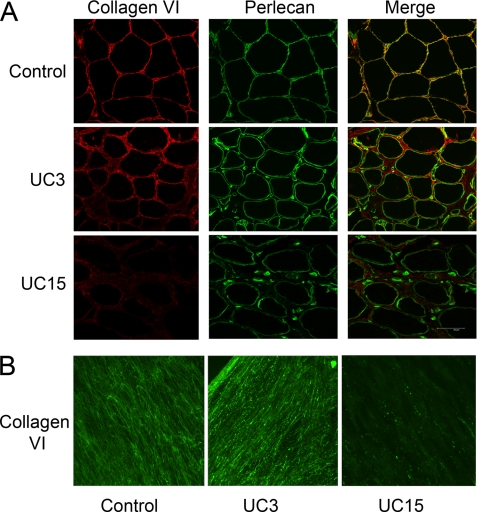
FIGURE 3.
Collagen VI immunostaining of muscle biopsies and dermal fibroblasts. A, cryosections of muscle biopsies from the control, UC3, and UC15 were immunostained with anti-collagen VI monoclonal antibody (red) and anti-perlecan polyclonal antibody (green). A merge of green and red fluorescence is shown. Collagen VI immunostaining was readily seen throughout the interstitial connective tissue of UC3 but significantly reduced in the UC15 muscle biopsies. Unlike the control muscle, collagen VI and perlecan did not co-localize in the basement membrane of the muscles from the patients. B, immunostaining of collagen VI deposited by fibroblasts from the control, UC3, and UC15. Cells were grown in the presence of 50 μg/ml ascorbate for 4 days post-confluency and then stained with the anti-collagen VI monoclonal antibody. Abundant but knotty collagen VI microfibrils were deposited by UC3 fibroblasts, whereas UC15 fibroblasts assembled few collagen VI microfibrils in the matrix.

