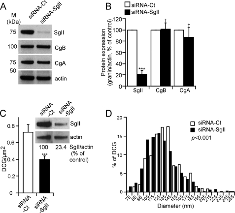
FIGURE 1.
Depletion and altered secretory granule morphology in response to siRNA-mediated silencing of SgII in wild-type sympathoadrenal PC12 cells. A, granins expression. Representative immunoblot of granins expression after siRNA silencing of SgII. Cell lysates from PC12 cells transfected with either siRNA-Ct or siRNA-SgII (96 h) were subjected to immunoblot detection of SgII, CgB, and CgA expression. Actin served as a normalization control. B, quantification of relative granin expression. The normalized SgII, CgB, and CgA expression in siRNA-Ct-treated cells was considered to be 100%. Values are given as the mean relative protein expression of seven independent experiments ± S.E. †, p > 0.05; ***, p < 0.001, t test. C and D, ultrastructural analyses of dense core granules after siRNA silencing of SgII. Micrographs from PC12 cells treated with siRNA-Ct or siRNA-SgII were analyzed for number of dense core granules per μm2 of cytoplasm (C) and diameter (nm) (D). n = 187 (siRNA-Ct) and 127 (siRNA-SgII) cell planes. C, immunoblot inset represents SgII expression before sample processing, quantified as described in B. ***, p < 0.001, Kruskal-Wallis test. D, dense core granule diameters were calculated from 572 (siRNA-Ct) and 586 (siRNA-SgII) randomly selected dense core granules. Values were ranked according to interval, and the distribution of the resulting populations was compared using a Kruskal-Wallis test.

