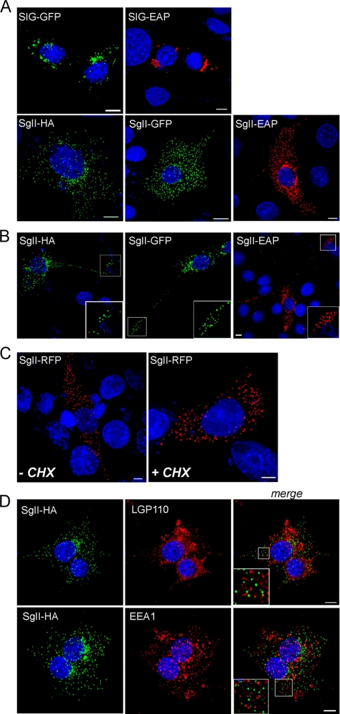
FIGURE 3.
Subcellular distribution of SgII fusion proteins expressed in neurosecretion-deficient sympathoadrenal A35C cells. A35C cells transfected (48 h) with expression plasmids encoding the indicated fusion proteins were processed for photoprotein fluorescence or immunocytochemistry and three-dimensional deconvolution microscopy. Shown are the three-dimensional views of 9–13 optical xy sections acquired along the z axis. Nuclei were visualized with Hoechst 33342 (blue). SIG-EAP, SgII-EAP (red), and SgII-HA (green) chimera were detected using the appropriate anti-reporter antibodies. Scale bars, 5 μm. A, cell bodies. B, neurites. Substantial accumulation of SgII-HA/GFP/EAP chimeric proteins is seen at the end of neurite-like structures, as exemplified in the enlarged insets of the deconvolved images. C, A35C cells expressing SgII-RFP were treated (3 h) with a mock buffer (DMSO; −CHX) or the protein synthesis inhibitor cycloheximide (CHX) (10 μg/ml; +CHX) prior to processing for photoprotein fluorescence. D, A35C-S7 cells (stably expressing SgII-HA) were co-stained for SgII-HA (green) and the lysosomal/late endosomal marker LGP110 or the early endosomal marker EEA1 (red). Absence of co-localization between SgII-HA and lysosomal or endosomal structures is exemplified in the enlarged insets from the merged three-dimensional images.

