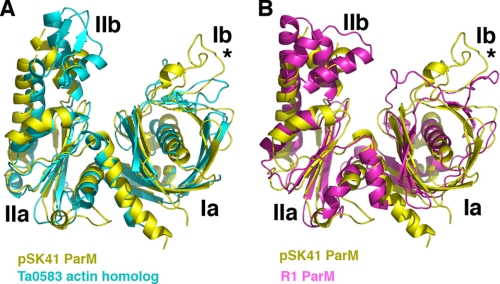
FIGURE 2.
Comparison of pSK41 ParM, R1 ParM, and Ta0853 structures. A, superimposition of the pSK41 ParM structure (yellow) onto the T. acidophilum Ta0583 actin-like protein structure (cyan). The structures overlay with a r.m.s. deviation of 2.4 Å for 83 corresponding Cα atoms. B, superimposition of the pSK41 ParM structure (yellow) onto the R1 ParM structure (magenta). The structures overlay with a r.m.s. deviation of 4.0 Å for 74 corresponding Cα atoms. The asterisks in both panels underscore that the loop region in Ib is the most structurally divergent region in all three structures.

