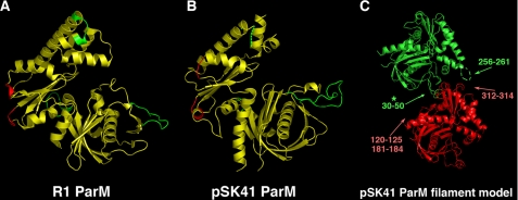
FIGURE 3.
Mapping of filament forming regions on ParM. A, regions involved in R1 ParM filament formation. The three key regions involved in intra-filament contacts are colored green and minor regions are colored red. B, mapping of the corresponding filament forming regions from R1 onto the pSK41 ParM structure. Key regions are green and minor regions are red as in A. C, pSK41 ParM filament model derived from the R1 ParM filament structure from Popp et al. (24). To produce the model, pSK41 structures were maximally docked onto the R1 structures. This figure underscores the large structural differences in the regions involved in filament contacts between R1 and pSK41 ParM proteins. Especially note that the large loop in pSK41 ParM subdomain Ib would clash with its interacting neighbor in such a filament, suggesting that pSK41 ParM might not form filaments similar in structure to R1 ParM (indicated by asterisk).

