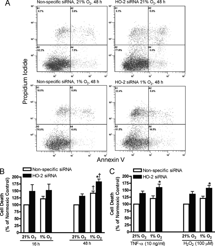
FIGURE 6.
A, representative annexin V/PI staining plots of HUVECs transfected with nonspecific or HO-2 siRNA exposed to 21% or 1% oxygen for 48 h. Cell death (% cells staining positive for Annexin V and/or PI) in HUVECs transfected with nonspecific or HO-2 siRNA exposed to 21% or 1% oxygen for 16 or 48 h (B) or exposed to 21% or 1% oxygen for 16 h in the presence of TNF-α or H2O2 (C). Bars represent means ± S.E. (n = 6 independent experiments); *, p < 0.05 for differences from nonspecific siRNA controls; †, p < 0.05 for differences from corresponding normoxic control values.

