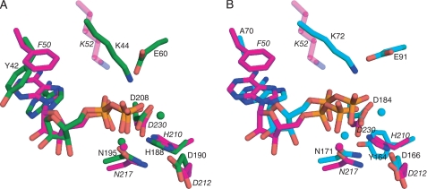
FIGURE 5.
Comparison of nucleotide binding in APH(9)-Ia, APH(3′)-IIIa, and protein kinase A. A, APH(9)-Ia, colored in magenta, and APH(3′)-IIIa, colored in green, and (B) APH(9)-Ia and protein kinase A (PDB code 1RDQ), colored in cyan, are superposed with the conserved amino acid residues to illustrate the orientation of the adenine ring in the nucleotide. The conserved residues and the nucleotides are shown in sticks and the magnesium ions are shown as spheres.

