FIGURE 1.
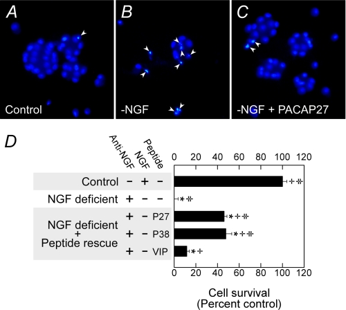
PACAP peptides promote sympathetic neuron survival after growth factor withdrawal. A–C, immature 5-day primary sympathetic neuronal cultures were withdrawn from NGF-containing serum-free medium for 24 h before fixation and Hoechst 33258 nuclear staining as described under “Experimental Procedures.” Neurons undergoing apoptosis after growth factor withdrawal (B) demonstrated nuclear condensation profiles (arrowheads), whereas cultures receiving 100 nm PACAP replacement presented far fewer nuclear apoptotic features (C). D, neuronal apoptosis after NGF deprivation was accentuated after 48 h as measured by MTS survival assays. Even under these circumstances, PACAP27 (P27) and PACAP38 (P38) demonstrated similar efficacy in protecting ∼50% of the population. VIP was less effective consistent with the prominence in PAC1 receptor expression in sympathetic neurons. Data represent a mean of 5–6 culture replicates for each treatment ±S.E. *, different from control; +, different from NGF-deficient cultures; ‡, different from VIP-treated cultures.

