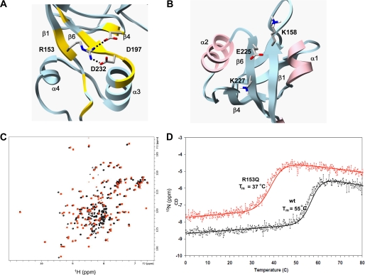
FIGURE 5.
Effects of human disease mutations on PDZ2+270 structure and stability. A, the structure of wild type PDZ2+270 shown with a hydrogen bond formed between positively charged guanidinium group of Arg153 and the negatively charged carboxylate group of Asp232 and Asp197. Residues with weighted backbone chemical shift difference greater than 0.1 ppm of mutant R153Q with reference to wild-type PDZ2+270 (supplemental Fig. S4C) are painted yellow. B, in wild type PDZ2+270, the negatively charged Glu225 is complemented by the positive charge of Lys158 and Lys227. C, overlay of 15N HSQC of PDZ2+270 (red) and mutant R153Q (black) at 30 °C. D, thermal unfolding curves of mutant R153Q (red) and wild-type PDZ2+270 (black) monitored by CD. The molar ellipticity (y axis) as a function of temperature (x axis) was fitted to a standard two-state unfolding equation.

