FIGURE 5.
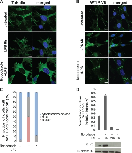
MT depolymerization impairs LPS-induced nuclear translocation of WTIP-V5. A, MT networks in GEC-WTIP-V5 cells. MT networks were visualized in GEC-WTIP-V5 cells in untreated control, LPS (1 μg/ml, 6 h), or nocodazole (10 μm, 20 min) followed by LPS treatment by immunostaining for FITC-tubulin. Scale bars, 8 μm. B, GEC-WTIP-V5 cells treated with LPS (1 μg/ml, 6 h), nocodazole + LPS (10 μm, 20 min), or untreated controls and fixed and immunostained for FITC-V5. Scale bars, 10 μm. C, quantification of WTIP-V5 accumulation. At least 150 cells from three separate experiments were imaged randomly and scored for localization of WTIP-V5. The bars represent the means ± S.E. (n = 3). D, nuclear extracts of GEC-WTIP-V5 cells were prepared from untreated control, LPS (1 μg/ml, 6 h or 24 h), or nocodazole (10 μm, 20 min) followed by LPS (1 μg/ml, 6 h). Detection of histone H3, a resident nuclear protein, was used as a nuclear loading control. Densitometric analysis was performed normalizing WTIP-V5 to histone H3 expression. Nuclear extracts of GEC-WTIP-V5 cells were prepared from untreated control, LPS (1 μg/ml, 6 or 24 h), or nocodazole (10 μm, 20 min) followed by LPS (1 μg/ml, 6 h). Detection of histone H3, a resident nuclear protein, was used as a nuclear loading control. IB, immunoblot.

