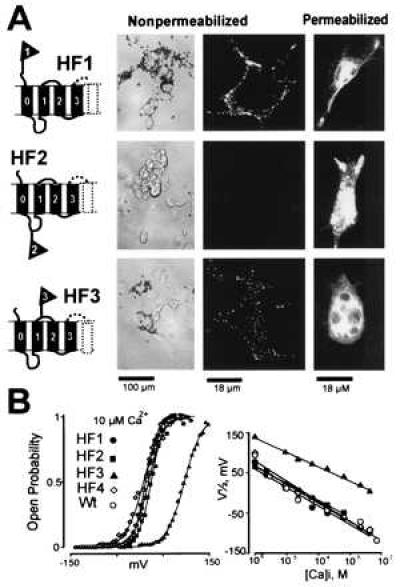
Figure 2.

MaxiK channels have an exoplasmic N terminus with an additional membrane spanning segment (S0). (A) Immunocytochemistry of c-myc tagged MaxiK channels (HF1, HF2 and HF3) expressed in COS-M6 cells. Cells were incubated with anti-c-myc mAb under nonpermeabilizing and permeabilizing conditions. Antibody binding was visualized using beads coated with secondary antibodies or with FITC-labeled secondary antibodies. Confocal images (two right panels) are from sections at the middle of the cells. Experiments were performed at least three times for each construct (also in Fig. 4) with similar results. (B) Functional expression of c-myc tagged clones in oocytes measured in inside-out patches. (Left) Voltage activation curves at 10 μM intracellular Ca2+, [Ca2+]i. Values for half activation potentials (V1/2) in 10 μM [Ca2+]i are (in mV): 12 ± 18 (n = 64) for wild-type Hslo (Wt); 5 ± 9 (n = 3) for HF1; 14 ± 8 (n = 6) for HF2; 98 ± 7 (n = 4) for HF3; 13 ± 6 (n = 3) for HF4 (see Fig. 4). (Right) V1/2 as a function of [Ca2+]i.
