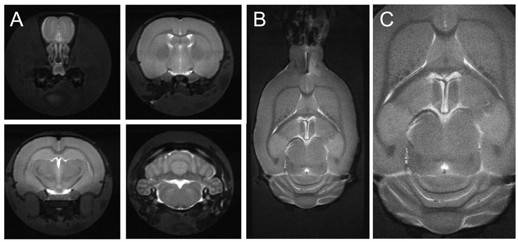
Figure 1.
Anatomical MR imaging on microwave-fixated rat brain. (A) Axial and (B/C) coronal T2-weighted, multi-slice, multi-shot RARE images (TR/TE = 3500/35 ms, 32 segments) of microwave-irradiated rat brain acquired over 750 µm and 600 µm thick slices, respectively. 256 × 256 and 640 × 320 data matrices were acquired for A and B/C, over 19.2 × 19.2 and 38.4 × 19.2 mm FOVs, respectively. Excellent soft-tissue contrast partially demonstrates structural integrity of microwave-irradiated samples. Data were acquired in 40 min (A) and 90 min (B/C). (C) represents an expanded view of (B).

