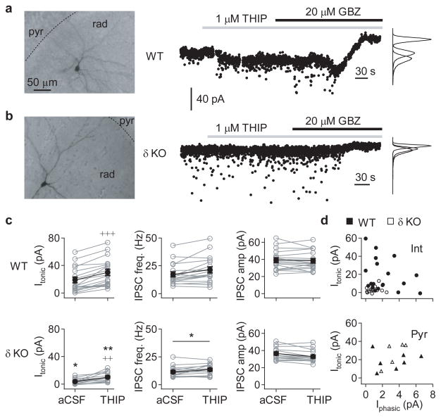
Figure 2. Hippocampal CA3 interneurons are disinhibited in adult Gabrd−/− mice.
GABAergic currents in hippocampal CA3 neurons were recorded in whole-cell voltage-clamp mode (Vhold = −70 mV) in the presence of 3 mM kynurenic acid, using a CsCl-based pipette solution. The pipette solution also contained biocytin, which allowed post-hoc classification of cell type based on morphology. (a) Representative recording from a CA3 radiatum interneuron (left) from a wildtype mouse. GABAergic currents were recorded during baseline conditions, and following bath application of 1 μM THIP (middle), and the tonic GABAergic current calculated relative to the holding current following bath application of 20 μM gabazine, or 50 μM picrotoxin, at the end of the experiment. (b) Representative recording from a similar CA3 radiatum interneuron (left) from a Gabrd−/− mouse, as in panel (a). (c) Summary of GABAergic currents recorded in CA3 interneurons from adult wildtype (top) and Gabrd−/− mice (bottom), showing the tonic GABAergic current (Itonic; left), frequency of spontaneous IPSCs (sIPSC; middle) and mean sIPSC amplitude (right), during both baseline conditions and following application of THIP. Error bars represent s.e.m. (d) Relationship between Itonic and the mean phasic current (Iphasic) in 1 μM THIP for CA3 interneurons (upper) and pyramidal neurons (lower). (*p < 0.05, **p < 0.01 cf. WT, +++p < 0.001, ++p < 0.01 vs baseline, Two-way repeated measures ANOVA followed by unpaired and paired t-tests, respectively)

