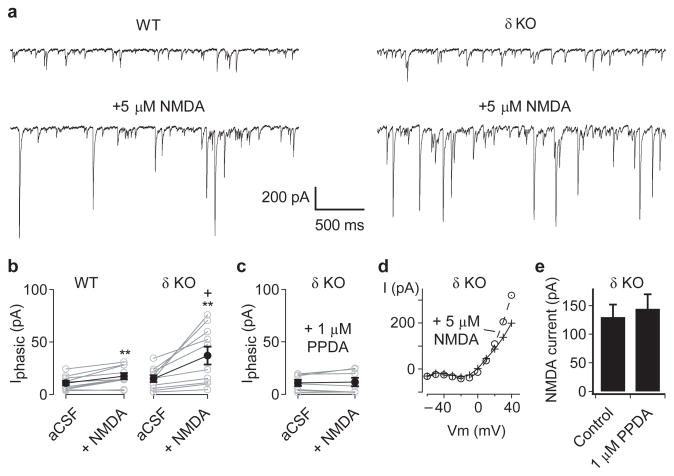
Figure 3. Hippocampal CA3 interneurons in adult Gabrd−/− mice are more sensitive to NMDAR-mediated excitation.
(a) To monitor the output of inhibitory interneurons, GABAergic currents were recorded from CA3 neurons pyramidal neurons, in the presence of 20 μM DNQX, using a CsCl-based pipette solution. Representative traces show the spontaneous IPSCs recorded during baseline conditions (upper) and following bath application of 5 μM NMDA (lower), in slices from adult wildtype (left) and Gabrd−/− mice (right). (b) Application of 5 μM NMDA increased the mean phasic current (Iphasic) in both wildtype (left) and Gabrd−/− mice (right), with significantly larger increases in inhibition in the Gabrd−/− mice. (c) The NMDA-evoked increases in Iphasic in Gabrd−/− mice were blocked by 1 μM PPDA. (d) To determine whether PPDA might act indirectly by blocking NMDAR on pyramidal neurons, recordings of NMDA currents were obtained in the presence of 20 μM DNQX, 20 μM gabazine and 5 μM ketamine, using a Cs-methylsulphonate based internal solution. The current-voltage plot before and after application of 5 μM NMDA, in a single pyramidal neuron, shows the voltage-dependent relief of ketamine block of the NMDAR at +40 mV (e) Pooled data, showing that 1 μM PPDA had no effect on the NMDA-evoked current recorded at +40 mV. (*p < 0.05 cf. WT, ++p < 0.01 vs baseline, Two-way repeated measures ANOVA followed by unpaired and paired t-test, respectively). Error bars represent s.e.m.

