
Figure 1.
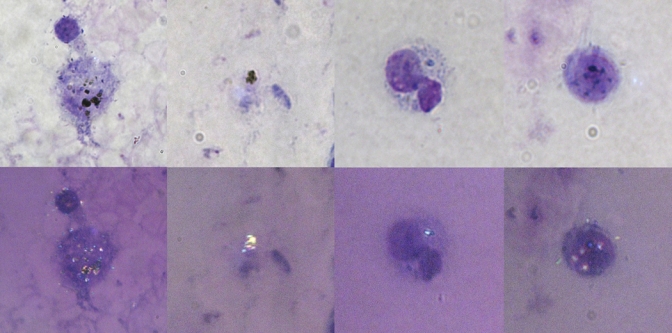
Plasmodium falciparum malaria pigment on a thick blood film from a patient with severe malaria. The upper panel shows pale images taken by using conventional light microscopy, and lower panel shows dark images taken by using the polarized light method described. This figure appears in color at www.ajtmh.org.