Figure 3.
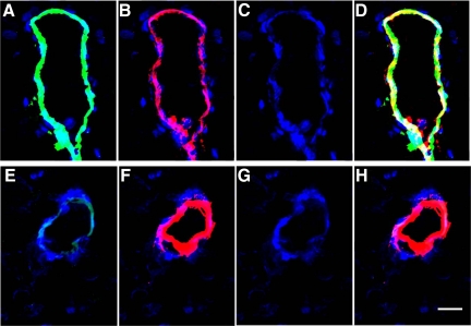
Alzheimer’s disease (A–D) and control (E–H) brain sections were examined by immunofluorescence for the presence of the endothelial cell marker Von Willebrand factor and for thrombin (×20). 4,6-Diamidino-2-phenylindole staining for nuclei was comparable in both AD (C) and control (G) sections. Staining for Von Willebrand factor was detectable in both AD (B) and control (F) tissues. In contrast, reactivity to the thrombin antibody (green immunofluorescence) was clearly present in AD (A) but barely detectable in control (E) vessels. Similarly, yellow/white immunofluorescence, which reflects a merged image of Von Willebrand and thrombin staining, was strong in AD sections (D) but not discernible in controls (H). Scale bar = 15 μm.

