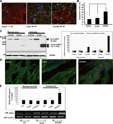
Figure 6.
CXCR4-overexpressed rats exhibit significant increase in cell death/apoptosis after ischemia-reperfusion in vivo. A: Increased cardiomyocyte susceptibility to ischemic injury was examined by assessing apoptosis using TUNEL assay. A double-staining technique was used (ie, TUNEL staining by using an In Situ Cell Death Detection Kit [Roche, USA] for apoptotic cell nuclei and DAPI staining for all cell nuclei. Representative images are shown. B: Apoptotic index was determined (ie, number of TUNEL-positive myocytes/total number of myocytes stained with anti-α-actinin × 100) from a total of 40 fields per heart. Assays were performed in a blinded manner. C: Increased cardiomyocyte susceptibility to ischemic injury was examined by assessing apoptosis using Western blot analysis of caspase 3 activation. Densitometric analysis of data from three different experiments is shown. *P < 0.05. D and E: The expression of inflammatory cytokine TNF-α was assessed in those hearts using immunofluorscence and QRT-PCR assay using a QuantiTect SYBR Green RT-PCR Kit. Specific primers for TNF-α and 18S were used. Primers were designed to generate short amplification products. Densitometric analysis of data from three different experiments is shown. *P < 0.05.

