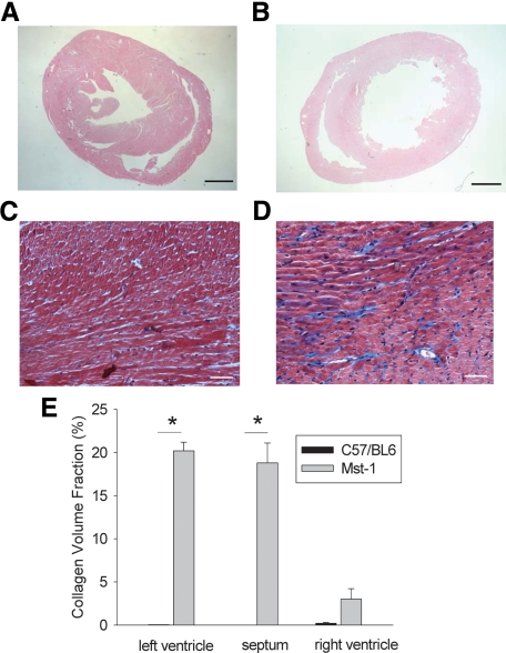
Figure 1.
A and B: Low-power photomicrographs of H&E-stained sections of C57BL/6 (A) and Mst1 transgenic (B) hearts from mice aged 13 weeks. Scale bars = 1 mm. C and D: Photomicrographs of Masson trichrome-stained sections of C57BL/6 (C) and Mst1 transgenic (D) mouse hearts. Scale bars = 50 μm. E: Bar graph representing collagen volume fraction in C57BL/6 and Mst1 transgenic mouse hearts. *P < 0.01.

