Figure 6.
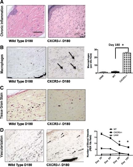
Recrudescence of an inflammatory-angiogenic response in persistent wound beds. A: Histopathological analysis of the acute and chronic inflammatory responses determined that there was a difference in macrophage or mononuclear leukocyte infiltration in the wounds of CXCR3−/− mice compared with wild-type mice. B: CXCR3−/− mice wound biopsies stained and quantified for increased macrophages in comparison with wild-type (WT) mice are shown. (Arrows indicate macrophages stained cells). C: Gram stain revealed no bacterial infection in both CXCR3−/− and wild-type mice. D: Excessive numbers of blood vessels persisted in the wounds of mice lacking CXCR3. Neovascularization in CXCR3−/− mice was assessed by using immunostaining of von Willebrand factor antigen outlining blood vessels of the mice. Representative von Willebrand factor immunostaining demonstrates the paucity of capillaries at day 180 in wild-type wounds compared with the CXCR3−/− wounds. Quantitative analyses of vascularization of CXCR3−/− mice wounds over time are shown in the graph. Representative micrographs are shown (n = 6 for each mouse genotype per time point). Scale bar = 50 μm. *P < 0.05.

