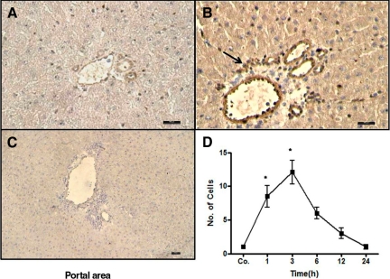
Figure 7.
Immunodetection of MCP-1/CCL2 in sham-irradiated sections (A) and 3 hours after irradiation (B). Sections were stained with a goat polyclonal antiserum directed against CCL2 followed by peroxidase staining. Increased expression of CCL2 was observed in the walls of the portal vessel and on some cells of the portal area (arrow). CCL2+ cells accumulated around the vessel with a peak at 3 hours as compared with sham-irradiated controls (original magnification, ×200; scale bar, 100 μm). C: Negative control was performed by using only secondary antibody against goat immunoglobulin followed by peroxidase staining (original magnification, ×100; scale bar, 100 μm). D: Counted CCL2+ cells in and around the portal field (N = 10). Results represent mean value of three animals and six slides per time point. Statistically significant at *P < 0.05 (mean ± SEM).

