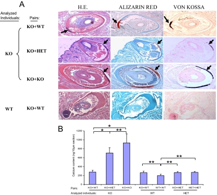
Figure 3.
The effect of parabiosis on aberrant mineralization of the connective tissue capsule of vibrissae. A: Two months after parabiotic surgery, the muzzle skin was biopsied and the mineralization was examined by H&E (left), Alizarin red (middle), and von Kossa (right) stains. The connective tissue capsule of vibrissae from the muzzle skin of KO mice paired with the wild-type (WT) mice developed less mineralization (the top panel, arrows) when compared with the KO mice paired either with KO (the third panel, arrows). Note that no mineralization was observed in the vibrissae of wild-type parabiotic mice in KO+wild-type pairing (bottom panel of A). B: Content of calcium in the vibrissae of KO mice in the different parabiotic pairs was determined by chemical assay. The calcium content was determined colorimetrically by using o-cresolphthalein complexone method, and the values are presented as mean ± SE. The total exact P value for the Kruskal-Wallis Test is 0.001; the exact P values for statistical comparison between KO mice in different groups are as follows: KO+wild-type versus KO+KO, 0.011; KO+wild-type versus KO+HET, 0.014; and KO+HET versus KO+KO, 0.242. Wild-type or HET in the pairs did not differ from the wild-type in wild-type+wild-type pair (*P < 0.05; **P > 0.05).

