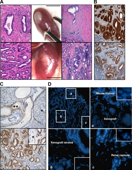
Figure 1.
Generation of mouse xenografts from primary localized human prostate tumors. Subrenal capsule xenografts of prostate tumor tissues (A, lower middle panel) often showed a robust vascularization that was not present in those derived from normal samples (A, upper middle panel). Hematoxylin and eosin staining of 5-micron FFPE sections of normal (A, upper right panel, ×200 magnification) and tumor (A, lower right panel, ×200 magnification) xenografts revealed that both maintain the histopathological features of the parental tissues (A, upper left and lower left panels, respectively). Tumor xenografts showed immunohistochemical markers of human PCa (PSA in B, upper panel, AMACR in B, lower panel, ×200 magnification; androgen receptor in C, lower panel, ×200 magnification, inset ×400). An example of Gleason 3 xenograft is shown. Scale bar = 10 mm. The xenografts did not show infiltration of mouse fibroblasts in the stroma, which was confirmed of human origin by both immunohistochemistry against the human androgen receptor (C, lower panel [asterisk represents human stromal cells]; C, upper panel, shows an orthotopic xenograft of human normal prostate as control [asterisk represents mouse prostate gland, double asterisk represents human prostate tissue]) and Hoechst staining (D; magnifications ×100 in the upper left panel, ×400 in the upper right and bottom panels, ×600 in the insets).

