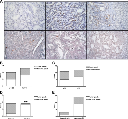
Figure 3.
Proliferation rate (percentage of Ki-67–positive nuclei) and apoptosis (percentage of Apoptag-positive nuclei) in parental prostate tumors and mouse xenografts. Tumors with higher proliferation rate (Ki-67 > 5%; A, upper middle panel, and D; **P < 0.01) grew preferentially in mice compared with those with low proliferation (Ki-67 < 5%; A, upper left panel). Representative examples of tumors with low and high apoptotic rates are reported in panel A, lower middle and lower left, respectively. Apoptotic rate, pathological stage, and GS did not affect the tumor take in mice (P > 0.05; B, C, and E). A, upper right and lower right, shows immunostaining of the xenografts derived from the tumors reported in A, upper middle and lower middle, which maintains the same proliferation rate and apoptosis of the parental tissues (×200 magnification).

