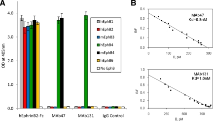
Figure 1.
Binding of monoclonal antibodies to EphB4. A: MAbs were immobilized on Protein A-Agarose beads and incubated with indicated recombinant EphB ECDs of human (h) or mouse (m) origin fused to AP. Negative controls included beads with AP alone (No EphB) and beads with unrelated IgG (IgG Control). As a positive control hEphrinB2-ECD fused to Fc was used in place of antibodies. B: To measure antibody affinity, biotinylated MAb47 (top) or MAb131 (bottom) were immobilized on Streptavidin-Agarose beads. Different concentrations (50 to 5000 pmol/L) of hEphB4-ECD-AP were applied to obtain saturation plot, nonspecific binding (no MAb added) values were subtracted and converted into coordinates of Scatchard plot.

