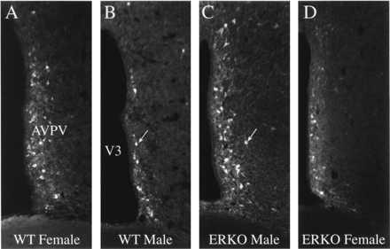
Figure 2.

TH expression in the AVPV of the ERKOα mouse. Images of immunohistochemically stained sections through the AVPV to illustrate the density and distribution of neurons that express TH in the AVPV of WT (+/+) female (A), WT (+/+) male (B), ERKOα male (C), or ERKOα female (D) mice. WT females have significantly more TH-immunoreactive neurons relative to that of WT males. Disruption of the ERα in the male (C) prevents the development of this sexually dimorphic pattern of TH expression. Cellular staining is noticeably reduced in the AVPV of the ERKOα female (D), but the number of TH-immunoreactive neurons is similar to that of WT females (see Fig. 3; ×200).
