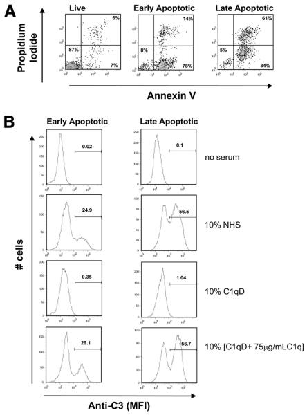
FIGURE 3.
C3b deposition on apoptotic cells. Early and late apoptotic Jurkats, as assessed by Annexin V/PI staining (A), were incubated with no serum, 10% NHS, 10% C1q-depleted serum (C1qD) (generated from the same NHS), or 10% [C1qD + 75 μg/ml C1q] for 30 min at 37°C and subsequently washed and probed with anti-C3 as described in Materials and Methods. C3 deposition was assessed by flow cytometry. Data shown are a typical histogram depicting the MFI from a single experiment, representative of three (B).

