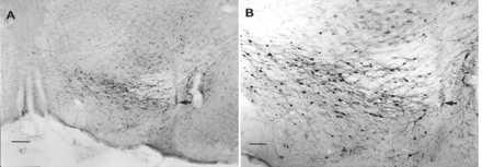
Figure 4.

Photomicrographs of nigral β-galactosidase expression 7 weeks after rAAV-CMV-lacZ injection. The nigral sections shown were taken from an rAAV-CMV-lacZ-injected rat that showed no evidence of a 6-OHDA lesion, and the rat was removed from the study. rAAV-CMV-lacZ-injected animals that were included in the study were found to have little or no β-galactosidase staining, probably because of the destruction of transduced cells by the subsequent striatal 6-OHDA lesion. (A) Low-power micrograph of the SN. The arrow indicates the injection site. (Bar = 200 μm.) (B) Higher-power micrograph of the same field. The arrow indicates the injection site. As has been observed (24, 41) most of the cells expressing β-galactosidase appear to be neurons. (Bar = 100 μm.)
