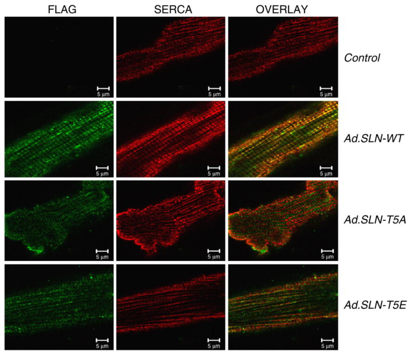
Fig. 1.

Confocal microscopic images of rat ventricular myocytes, showing co-localization of WT and mutant SLN with SERCA2a. Adult rat ventricular myocytes were infected with Ad. WT-SLN, Ad.SLN-T5A or Ad.SLN-T5E or left uninfected and stained with FLAG antibody (green) and SERCA2a (red) antibody. Orange-overlay of images shows co-localization.
