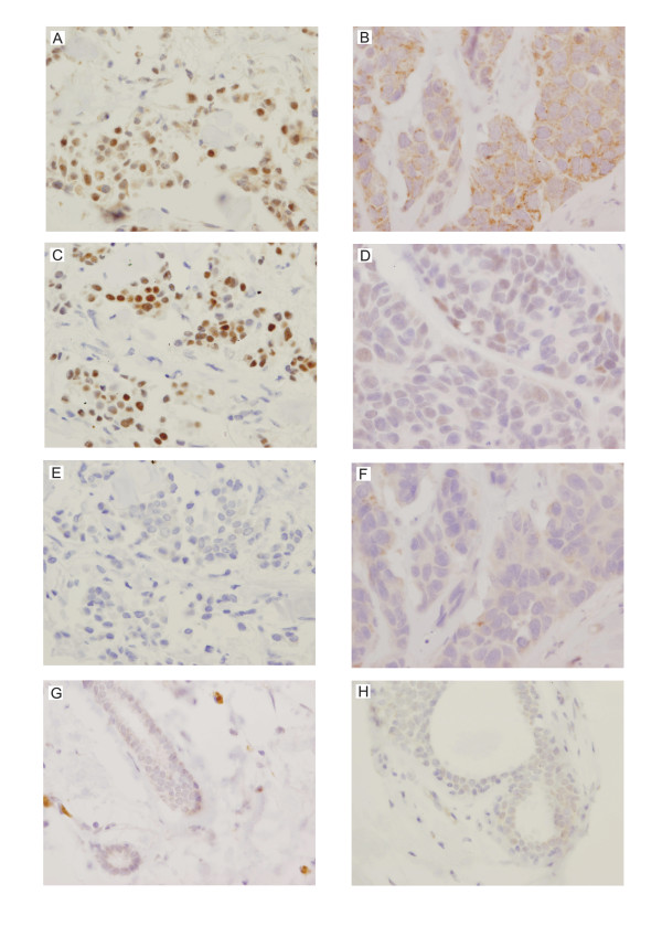
Figure 7.
Representative immunohistochemical staining of the analyzed breast cancer samples shows elevated levels of HAX-1 in tumor tissues. Each column represents samples from one patient. HAX-1 staining was detected in the nuclei (A) and the cytoplasm (B) of tumor cells (magnification × 400). Nuclear HAX-1 localization was observed only in the strongly ER-positive samples (C), while in the samples with cytoplasmic HAX-1 staining ER expression was weak (D). Control samples incubated with mouse IgG of the same subclasses and concentrations as the primary antibody (E and F) were negative. Normal samples from the analyzed two patients (G and H) do not show visible HAX-1 staining.

