Figure 6.
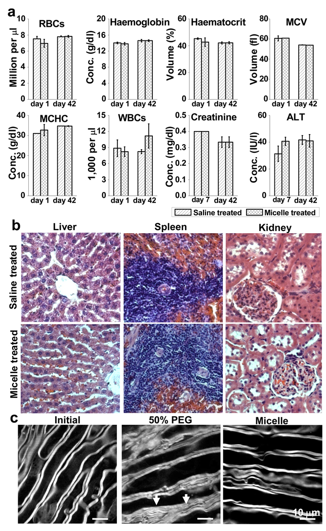
Toxicity analysis. a, Following injection of 1 mL mPEG-PDLLA micelle or saline solution, neither complete blood count of Long-Evans rats at day 1 and day 42 or serum analysis at day 7 and day 42 shows adverse effects between the two groups. b, Histological analysis of explanted liver, spleen and kidney with haematoxylin and eosin staining between the control group and the micelle treated group indicates no signs of cellular or tissue damage. Magnification: 400×. c, Morphological analysis. After incubating a healthy spinal cord strip (left) in 50% PEG in Krebs’ solution for 17 min, the axons became attached to each other (middle). The white arrows indicate possible fusion of adjacent axonal myelin. In contrast, after incubation with 0.67 mg/mL mPEG-PDLLA micelles for 180 min (right), a spinal cord strip displays no obvious morphological changes of myelin (right).

