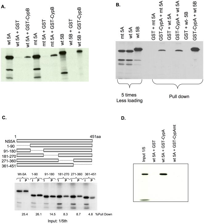
Figure 5. NS5A binds both CypB and CypA.
(A) wt-NS5A, mt-NS5A, and wt NS5B-FLAG (1bN) were labeled with 35S methionine/cysteine by in vitro translation. Labeled proteins were incubated with either GST alone (Lanes 2, 5, and 8) or GST-CypB (Lanes 3, 6, and 9). Gluthathione Sepharose beads were used to purify GST-containing complexes from the binding mixture. After separation via SDS-PAGE, 35S-labeled proteins were detected by autoradiography. Lanes 1, 4, and 7 contain labeled proteins used in the binding assay. (B) The wt-NS5A, mt-NS5A, and wt NS5B-FLAG (Con 1b) were in vitro translated and incubated with either GST alone or GST-CypA. The GST binding assay was performed as described in (A). The left three lanes contain labeled proteins used in the binding assay. (C) The binding region within the NS5A region for CypA was mapped by generating 90 amino acids in-frame deletion mutants. The radiolabeled mutant proteins were incubated with GST-CypA as above. Input protein (I) was loaded at 1/5 volume alongside the corresponding pull-down product (P). Pulled down signal was quantified using Storm and % values of pull down are shown for each deletion mutant. (D) The CypA containing mutations in the isomerase active site was analyzed for binding to wt-NS5A using GST binding assay.

