
Figure 2. Contribution of CMA dysfunction to pathogenesis in neurodegeneration.
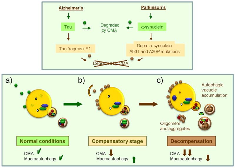
Different pathogenic proteins (α-synuclein and Tau) depicted here, have been shown to have a primary detrimental effect on CMA activity. Whereas wild-type or unmodified forms of these proteins (green circles) can be targeted and taken up by lysosomes via CMA (a), their pathogenic counterparts (brown circles) are delivered to the CMA translocation complex at the lysosomal membrane but fail to translocate into the lumen (b). These pathogenic proteins often organize into irreversible oligomeric complexes at the lysosomal membrane that further block CMA activity. Cells respond to CMA blockage by upregulating macroautophagy, the only proteolytic pathway that can directly degrade proteinaceous inclusions and aggregates (b). Failure of macroautophagy with time – due to exhaustion or primary damage by the pathogenic proteins – could precipitate progression of disease (c).