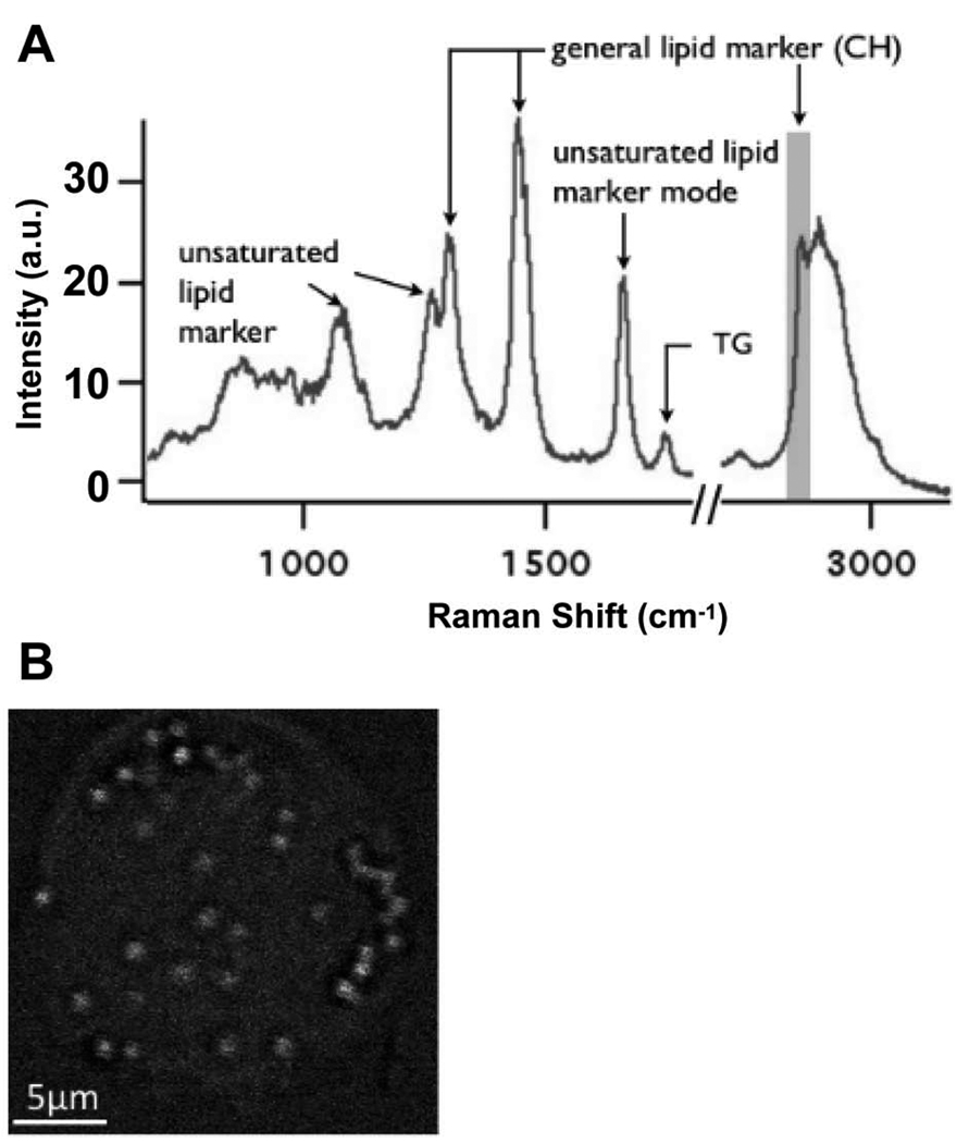
Figure 2. CARS imaging of THP-1 monocytes confirms the lipid nature of the droplets.
After a 3-hour treatment with VLDL + LpL (200 mg TG/mL, 2 U/mL), THP-1 monocytes were analyzed and imaged by CARS microscopy. (A) Raman spectra obtained from individual lipid droplets inside monocytes exhibit all the hallmarks of lipids. (B) A chemical image obtained by utilizing the general lipid marker mode of an aliphatic CH vibration at 2845 cm−1 to generate the contrast confirms that the droplets inside monocytes are highly enriched in lipids. Lipid droplets are ~1 µm in diameter throughout the cell. Images and spectra were obtained on home-built instruments utilizing and Olympus I×71 inverted microscope as platform, equipped with an Olympus Plan-Apo 100×, 1.4 NA objective lens. Cells were fixed with 2% paraformaldehyde, washed in phosphate buffered saline, and imaged at room temperature using Picoquant SymPhoTime software. n = 3; each replication represents the analysis of 5–8 fields of view. A.u., arbitrary units.

