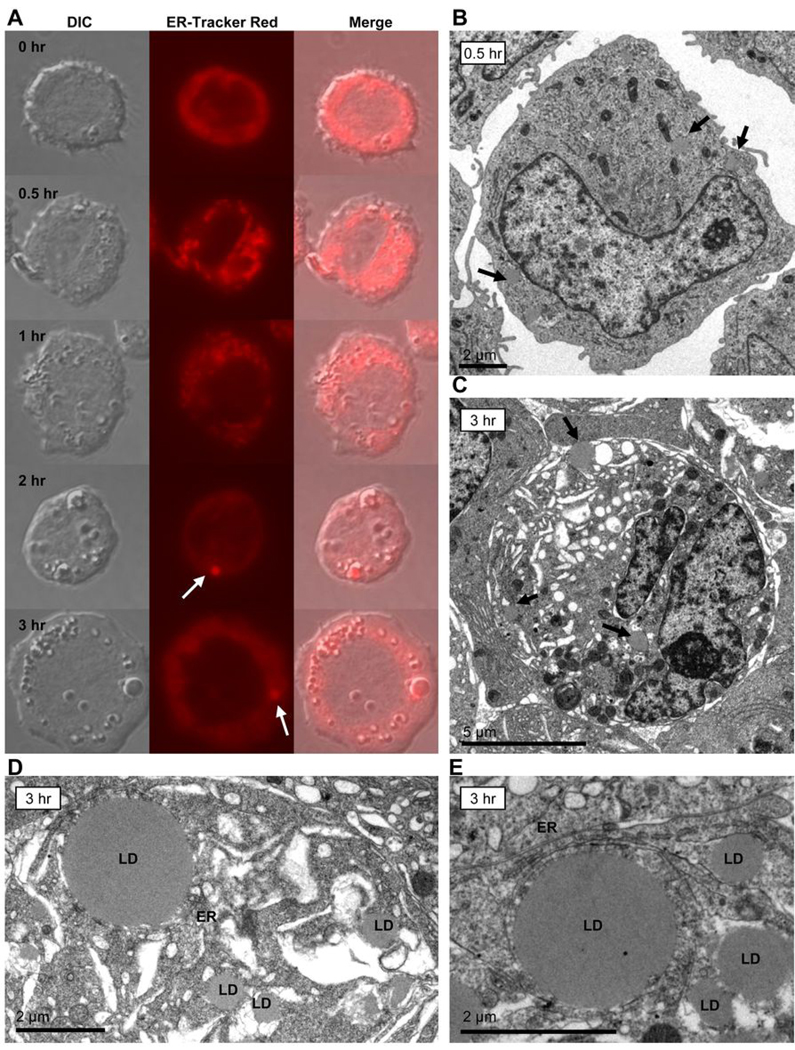
Figure 3. VLDL lipolysis product-induced lipid droplets are found in close proximity to the ER.
(A) THP-1 monocytes were treated at a cell density of 1 × 10⁶ cells/mL with VLDL + LpL (200 mg TG/dL, 2 U/mL) over a time course ranging from 0.5 to 3 hours. At each time point cells were collected and stained with ER-Tracker Red to observe any interactions between ER structures and lipid droplet formation. Monocytes were observed using a Zeiss Axioskop2 plus microscope with a Pan-Neofluar 40× objective, 1.3 oil. Images were captured using a Zeiss AxioCam MRm camera and processed using AxioVisionLE software. Fluorescence and DIC images of the same cells are shown. At 2 hours, ER-associated contents are seen in the same area as some of the lipid droplets within the cell (ER-Tracker Red, arrow). By 3 hours, the ER appears to surround the lipid droplet and/or its contents (arrow). Cells from each treatment were analyzed over a minimum of 10 frames per experiment. The experiment was repeated 3 times, with similar results. (B to E) TEM of THP-1 monocytes treated with VLDL + LpL (200 mg TG/dL, 2 U/mL) for either 0.5 or 3 hours. Monocytes were harvested, prepared for TEM analysis, and embedded in epoxy resin for ultra thin sectioning. The images were collected on a Phillips CM120 microscope at 80kV using a Gatan MegaScan model 794/20 digital camera. (B) Monocyte treated with VLDL lipolysis products for 0.5 hours contains small, disperse lipid droplets (original magnification ×8510, arrows). (C) Monocyte treated with VLDL lipolysis products for 3 hours contains larger lipid droplets (original magnification ×8510, arrows). (D and E) Higher magnification images of monocytes treated with VLDL lipolysis products for 3 hours reveal lipid droplets of multiple sizes (D, original magnification ×15900) and lipid droplets in close proximity to the ER (E, original magnification ×11000). LD, lipid droplet; ER, endoplasmic reticulum.

