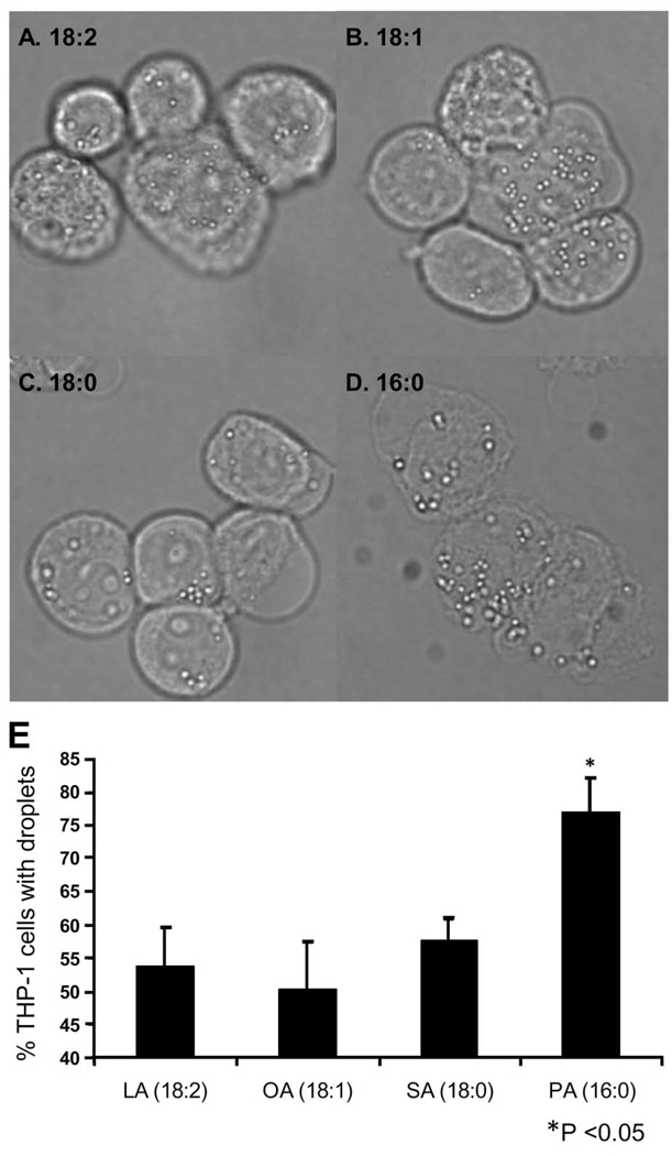
Figure 5. Synthetic fatty acids induce formation of lipid droplets in THP-1 monocytes.
THP-1 monocytes were treated at a cell density of 1 × 10⁶ cells/mL with linoleic acid (A, LA (18:2), η-6), oleic acid (B, OA(18:1)), stearic acid (C, SA(18:0)), or palmitic acid (D, PA(16:0)) at final concentrations of 150 µM for 3 hours. Monocytes were observed by phase contrast microscopy using an Olympus B×41 microscope with a 60× objective, NA 0.80. Images were captured using an Olympus QColor3 camera and processed using QCapture software. A minimum of 300 cells were counted from each treatment, and the average percentage of cells positive for lipid droplets is represented in panel E (n = 3, *P<0.05). Original magnification ×600 for all panels.

