
Figure 3b:
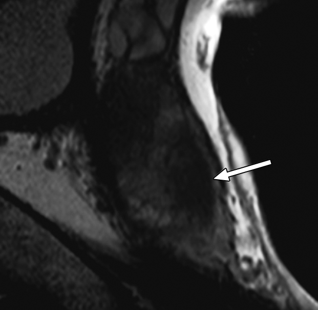
Prostate cancer in 58-year-old man. (a) Axial and (b) sagittal T2-weighted MR images demonstrate a low-signal-intensity lesion (arrow) in left mid PZ suspicious for cancer. (c) MR spectra demonstrate increased ratio of choline (cho) to citrate (cit) in left mid PZ lesion (∗) when compared with normal right side. (d) Raw dynamic contrast-enhanced MR image and (e) Ktrans and (f) kep maps help localize tumor (arrow). (g) Histopathologic slide at mid prostate level confirms presence of tumor (Gleason score, 7), with extracapsular extension (red line) detected on MR images. A = anterior, L = left, P = posterior, R = right.