
Figure 4c:
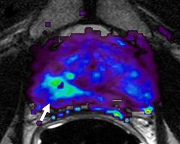
Prostate cancer in 62-year-old man. (a) Axial T2-weighted MR image demonstrates a low-signal-intensity focus (arrow) at right apex mid PZ suspicious for prostate cancer. (b) Raw dynamic contrast-enhanced MR image and (c) Ktrans and (d) kep maps help localize tumor (arrow). (e) Histopathologic slide at apex mid prostate level confirms presence of tumor (Gleason score, 8) more anteriorly (red line), secondary to distortion and shrinkage of specimen. Keys are same as for Figure 3.