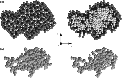
Figure 11.
Representation of methionine-rich 2S albumin atomic structure and of its solvation dummy spheres found by SASMOL. (a) View of tetrahedral close-packed spheres assigned to the protein (white), the first solvation shell (black) and the second solvation shell (grey). (b) Spacefill representation of the protein's atoms. In the left column, all tcp spheres and atoms are shown, whereas in the right column only the ones with the coordinates z < 0 are reported.

